| Table of Contents |  |
|
Clinical Image
| ||||||
| Cardiac metastasis mimicking myocardial ischemia | ||||||
| Laura Evangelista1, Gentian Denas2, Alessandra Bianchi2, Anna Rita Cervino1, Alberto Banzato2 | ||||||
|
1Radiotherapy and Nuclear Medicine, Veneto Oncologic Institute, IRCCS, Padua, Italy.
2Cardiology Unit, Veneto Oncologic Institute, IRCCS, Padua, Italy. | ||||||
| ||||||
|
[HTML Abstract]
[PDF Full Text]
[Print This Article]
[Similar article in Pumed] [Similar article in Google Scholar] |


| How to cite this article |
| Evangelista L, Denas G, Bianchi A, Cervino AR, Banzato A. Cardiac metastasis mimicking myocardial ischemia. Edorium J Cardiol 2015;2:1–3. |
|
Case Report
|
|
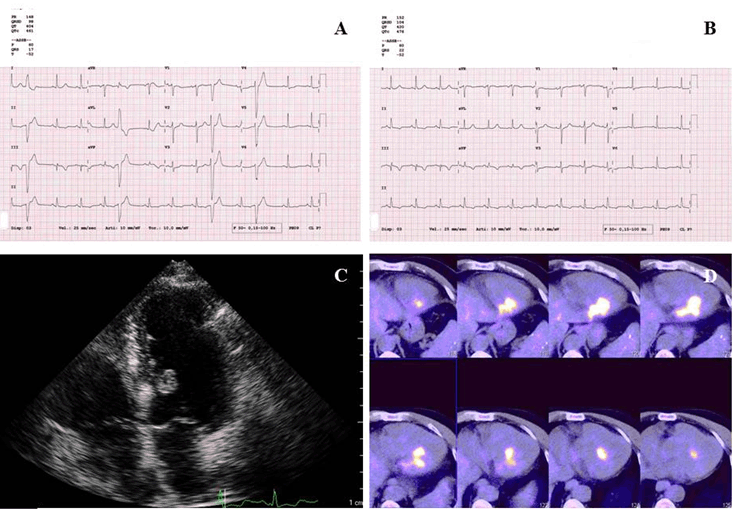
A 74-year-old male, with a history of follicular thyroid carcinoma since 2004, already treated by surgery and serial radioiodine therapies, was referred to a cardiologic evaluation because of extra systolic beats and ischemic T waves in inferior leads showed in resting electrocardiogram (ECG), that was performed due to zoledronic acid assumption. The patient was asymptomatic showing normal values of troponin-I. A transthoracic echocardiography revealed a mass arising from the inter-ventricular septum and protruding into left ventricle. In order to identify the nature of cardiac mass a whole-body 18F-fluorodeoxyglucose (FDG) positron emission tomography (PET)/computed tomography (CT) scan was performed. PET/CT images demonstrated an intense FDG-uptake in the septum and in left ventricular cavity. In Figure 1 shows all collected images from the patient. A biopsy obtained through mediastinoscopy, and subsequent histological analysis confirmed a cardiac metastasis from follicular thyroid cancer. Due to the rapid progression of disease, the patient died after six months. Therefore, none specific therapy was started for the cardiac metastasis. |
|
|
|
Discussion
|
|
In chronically stable cancer, patients without any cardiac symptoms suggestive of ischemia, an electrocardiogram pattern of myocardial ischemia should raise the suspicion of cardiac metastasis. Although cardiac metastasis are identified in less than 1% of patients who die of thyroid cancer [1], the appearance of symptomatic angina in such patients should be carefully evaluated. Cardiac magnetic resonance and non-ECG-gated multi-detector CT with intravenous contrast provide adequate information of the cardiac mass extension [2] [3]. FDG PET/CT can be alternatively used in case of a negative 131I-uptake on scintigraphy scan [4]. A lot of cardiac masses have been already described by FDG PET/CT [5] [6] [7], but in the majority of cases they arise in the atrium (such as angiosarcomas, atrial myxoma) or in the interatrial septum (such as massive fatty deposit, lipomatous hamartoma and similar). However in few cases, they can arise in the ventricular cavity and therefore should be considered for the differential diagnosis. |
|
Conclusion
|
|
The discovery of a cardiac metastasis in an oncological patient, may change both therapeutic management and prognosis. Keywords: Cancer, Cardiac metastasis, Radioiodine, Thyroid |
|
References
|
|

|
[HTML Abstract]
[PDF Full Text]
|
|
Author Contributions
Laura Evangelista – Substantial contributions to conception and design, Analysis and interpretation of data, Drafting the article, Final approval of the version to be published Gentian Denas – Substantial contributions to conception and interpretation of data, Drafting the article, Final approval of the version to be published Alessandra Bianchi – Substantial contributions to conception and interpretation of data, Drafting the article, Final approval of the version to be published Anna Rita Cervino – Substantial contributions to interpretation of data, Drafting the article, Final approval of the version to be published Alberto Banzato – Substantial contributions to conception and design, Analysis and interpretation of data, Drafting the article, Final approval of the version to be published |
|
Guarantor of submission
The corresponding author is the guarantor of submission. |
|
Source of support
None |
|
Conflict of interest
Authors declare no conflict of interest. |
|
Copyright
© 2015 Laura Evangelista et al. This article is distributed under the terms of Creative Commons Attribution License which permits unrestricted use, distribution and reproduction in any medium provided the original author(s) and original publisher are properly credited. Please see the copyright policy on the journal website for more information. |
|
|