| Table of Contents |  |
|
Case Report
| ||||||
| Long inflation to left main coronary artery with kissing balloon inflation by drug-coated balloon and perfusion balloon | ||||||
| Keisuke Nakabayashi1, Hisayuki Okada1, Hideki Saito1, Toshiaki Oka1 | ||||||
|
1Department of Cardiology, Seirei Hamamatsu General Hospital, 2-12-12 Sumiyoshi, Naka-ku, Hamamatsu City, Shizuoka 430-8558, Japan.
| ||||||
| ||||||
|
[HTML Abstract]
[PDF Full Text]
[Print This Article]
[Similar article in Pumed] [Similar article in Google Scholar] |


| How to cite this article |
| Nakabayashi K, Okada H, Saito H, Oka T. Long inflation to left main coronary artery with kissing balloon inflation by drug-coated balloon and perfusion balloon. Edorium J Cardiol 2016;3:1–5. |
|
Abstract
|
|
Introduction:
The advancement in innovation of devices and techniques has allowed percutaneous coronary intervention to be used in increasingly complex cases. However, in-stent restenosis involving a bifurcation lesion remains a problem in the drug-eluting stent era. Drug-coated balloons that administer drugs to vessel walls are effective in drug-eluting stent in-stent restenosis even at bifurcation lesions. It might be incompatible with proximal lesions because of the ischemia induced by the long inflation required for drug-coated balloon (approximately 30–60 sec).
Case Report: We described a 77-year-old male suffering from frequent in-stent restenosis of the left circumflex ostium that had been treated with a conventional balloon and a scoring balloon that has element to crack vessel wall. We treated this case with the novel approach consisting of long kissing balloon inflation at the left main coronary artery with a drug-coated balloon and perfusion balloon that guarantees the blood flow from left main to left descending artery, thereby preventing overt ischemia. He has been free from symptoms eight months after the procedure. Conclusion: Perfusion balloon could be a helpful option when we treat the in-stent restenosis of proximal and bifurcated lesion by a drug-coated balloon. | |
|
Keywords:
Drug-coated balloon, Kissing balloon inflation, Left main trunk, Perfusion balloon
| |
|
Introduction
| ||||||
|
The advancement in innovation of devices and techniques has allowed percutaneous coronary intervention (PCI) to be used in increasingly complex cases. However, in-stent restenosis (ISR) involving a bifurcation lesion is still a problem in the drug-eluting stent (DES) era. Drug-coated balloons (DCB) are effective in DES-ISR [1] even at bifurcation lesions [2] [3]. The application of the technique for ostial lesions, especially in the left main coronary artery (LMCA), is difficult because of the ischemia induced by the long inflation required for DCB (approximately 30 seconds). Here we describe a novel approach for managing the ISR of left circumflex (LCX) ostial lesion, consisting of the use of a long kissing balloon inflation (KBI) by DCB and perfusion balloon (PB) without overt ischemia. | ||||||
|
Case Report
| ||||||
|
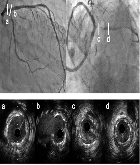
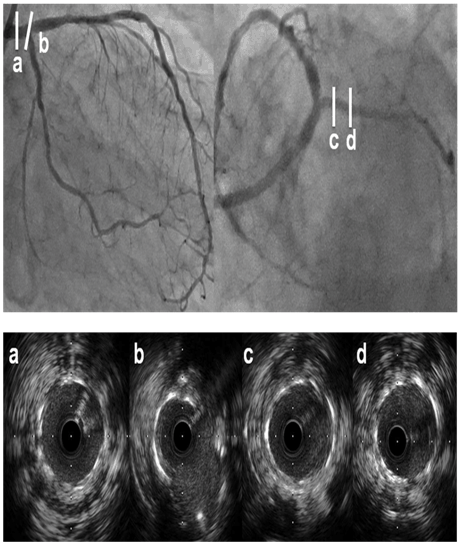
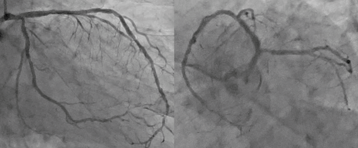
A 77-year-old male with hypertension, type 1 diabetes mellitus, and stage 3 chronic kidney disease underwent PCI three times. Firstly, because of effort angina pectoris, we treated three vessels using a biolimus eluting stent (3.0×14 mm Nobori, Terumo, Tokyo, Japan) from the proximal right coronary artery, 3.0×24 mm Nobori to proximal left descending artery (LAD). In staged, we deployed an everolimus-eluting stent (2.5×33 mm Xience Prime, Abbott Vascular Devices, Redwood City, USA) from LMCA to just proximal of the LCX lesion and subsequently performed KBI at the LMCA bifurcation. The patient suffered a recurrent angina eight months after the procedure. Angiography showed ISR of the LCX ostium, and we subsequently dilated the ISR lesion with a conventional non-compliant balloon (3.5×20 mm Powered Lacrosse2, GOODMAN, Aichi, Japan). This dilation induced carina shift to the LAD; therefore, we deployed a 3.5×18 mm Xience Prime from the LMCA to just proximal of the LAD and added KBI as if "staggered" culottes stenting. Final KBI caused the overt ischemia, despite of the short inflation time. Unfortunately, the patient suffered a recurrent angina six months after the second procedure, and angiography showed a similar ISR. As a treatment regime, the lesion was dilated with a scoring balloon (3.5×13 mm Lacrosse NSEa, GOODMAN, Aichi, Japan) and KBI was added. The patient again suffered from recurrent angina six months after the third procedure. He had been taking 100 mg of aspirin and 75 mg of clopidogrel daily since first PCI. Angiography showed most of the same ISR of the LCX ostium (Figure 1). A coronary artery bypass graft procedure was offered. However, the patient refused and instead elected for PCI. Therefore, we recommended the use of a DCB, to which he agreed. Written informed consent was obtained from the patient. We passed a 7Fr-guiding catheter (VL 3.5SH, Boston Scientific, Massachusetts, USA) to the left coronary artery via the right radial artery and then advanced two soft wires (SION and SION blue, ASAHI INTECC, Aichi, Japan) to the LAD and LCX. Intra-vascular ultrasound study (IVUS, Opticross, Boston Scientific, Massachusetts, USA) showed no stent malposition or fractures but did show neointimal hyperplasia (Figure 1). We prepared a PB (3.5×20 mm Ryusei, Kaneka Medix, Osaka, Japan) proximal to the LAD for KBI, which prevented ischemia of the LAD perfusion area and carina shift. Pre-dilation was performed with KBI at the LMCA-LCX with a 3.5×15 mm Powered Lacrosse2 followed by a 3.5×13 mm Lacrosse NSEa at the LMCA-LAD with a 3.0×20 mm Ryusei. There was no sign of ischemic change detected on the electrocardiogram. Finally, we performed KBI with a DCB (3.5×20 mm SeQuent Please, B Braun, Melsungen, Germany) at the LMCA-LCX and a 3.0×20 mm Ryusei at the LMCA-LAD. Coronary blood flow to the LAD was confirmed (Figure 2). Inflation time was 60 seconds. Despite a 1-mm ST depression in the inferior leads, the hemodynamic change and chest pain did not reappear. The results from a subsequent angiography and IVUS were considered acceptable (Figure 3). This patient was free from ischemic symptoms eight months after the procedure and had no ISR at the re-study coronary angiography (Figure 4). | ||||||
|
| ||||||
|
| ||||||
|
| ||||||
| ||||||
|
Discussion
| ||||||
|
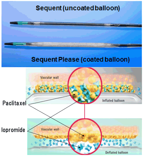
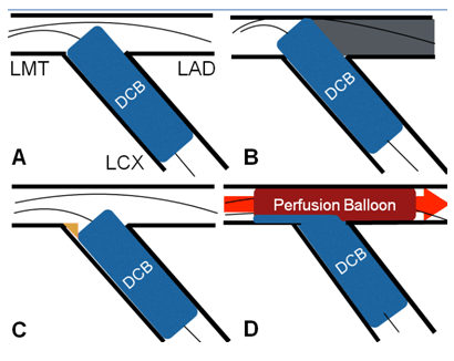
The patient was treated with a DCB after a third attempt with ISR of the LCX ostium that was previously treated with a conventional balloon and a scoring balloon. However, the long inflation time of the DCB to the LCX ostium (regardless of whether KBI was performed or not) could potentially induce severe ischemic complications because of total left coronary artery area ischemia. Therefore, we elected to perform the KBI to the LMCA lesion, guaranteeing blood flow by the PB from the LMCA to the LAD. Bifurcation lesions remain a challenging area in the DES era, and this case had a high frequency of ISR. Notably, the LCX ostium had a high ratio of ISR irrespective of the LMCA-LCX or LMCA-LAD stenting because of the acute angle and hinge motion of the origin of the LMCA [4]. Nevertheless, several methods (e.g., repeat stenting with DES, DCB, scoring balloon or conventional balloon angioplasty alone) were attempted to overcome the lesions because the existing strategies were indeterminate. One study reported that patients treated with paclitaxel-eluting stents had 25% of recurrent binary restenosis. Furthermore, 27% of those treated with a paclitaxel-eluting balloon and 57% of those treated with balloon angioplasty alone had ISR of limus-eluting stent [1]. Another study reported that the use of a paclitaxel-eluting balloon was associated with favorable rates of major adverse cardiac events and target lesion revascularization at a 1-year follow-up compared with the implantation of an everolimus-eluting stent [5]. Furthermore, DES re-implantation caused several stent layers in the coronary vessel wall and could negatively affect the long-term outcome [6]. DCB angioplasty offers an effective treatment for DES-ISR without the necessity of implanting additional metal layers for drug release [7] and was concluded to be potentially beneficial. However, DCB is a device to administer the drug to the vessel wall and requires a long inflation time of approximately 30 sec (Figure 5). DCB and LMCA lesions seemed to be incompatible. If we can position DCB precisely just proximal to the ostium, DCB alone may be effective. Because DCB has to be positioned only one time, it is too difficult. If DCB were positioned more proximally, it would lead to overt ischemia. If DCB were positioned more distally, it would lead to residual stenosis (Figure 6A-C). The perfusion balloon guarantees the distal flow. The DCB's merit is its relatively high patency and ability to avoid additional stents. DCB's demerit is inducible ischemia due to required long inflation time and one shot deal. Perfusion balloon might cancel out the demerit (Figure 6D). Perfusion balloon was designed to provide continuous transcatheter blood flow and thereby reduced myocardial ischemia during coronary angioplasty (Figure 7). Perfusion baloon was used for a long inflation balloon angioplasty especially for ISR and for small vessels, thrombus control, and the prevention of acute perfusion injury of acute myocardial infarction. Perfusion baloon had gone out of production in 2008 in Japan because of its unprofitability but has been revived in response to interventionists' willingness. Recently, PB was often used during complicated situations such as coronary dissection or perforation. We could perform KBI at the LMCA lesion in a relatively safe manner with PB guaranteeing continuous blood flow from the LMCA to the LAD. Nonetheless, the electrocardiogram showed that ischemic change originated in the LCX occlusion, and we could avoid the critical ischemia complication despite the long inflation time for KBI at the LMCA lesion. To the best of our knowledge, the use of Perfusion baloon for this purpose has not been reported, and the use of KBI by DCB and PB may be a useful option in practice. | ||||||
|
| ||||||
|
| ||||||
|
| ||||||
|
| ||||||
|
Conclusion
| ||||||
|
We presented the case treated with perfusion balloon and drug-coated balloon (DCB) for recurrent ISR of the left circumflex artery (LCX) ostium. The DCB is an attractive device for ISR; however, it may be incompatible in cases with left main coronary artery (LMCA) ostial lesions. In these cases, the additional use of perfusion balloon may compensate for this incompatibility. | ||||||
|
References
| ||||||
| ||||||


|
[HTML Abstract]
[PDF Full Text]
|
|
Author Contributions
Keisuke Nakabayashi – Substantial contributions to conception and design, Acquisition of data, Analysis and interpretation of data, Drafting the article, Revising it critically for important intellectual content, Final approval of the version to be published Hisayuki Okada – Substantial contributions to conception and design, Acquisition of data, Analysis and interpretation of data, Revising it critically for important intellectual content, Final approval of the version to be published Hideki Saito – Substantial contributions to conception and design, Acquisition of data, Analysis and interpretation of data, Revising it critically for important intellectual content, Final approval of the version to be published Toshiaki Oka – Substantial contributions to conception and design, Acquisition of data, Analysis and interpretation of data, Revising it critically for important intellectual content, Final approval of the version to be published |
|
Guarantor of submission
The corresponding author is the guarantor of submission. |
|
Source of support
None |
|
Conflict of interest
Authors declare no conflict of interest. |
|
Copyright
© 2016 Keisuke Nakabayashi et al. This article is distributed under the terms of Creative Commons Attribution License which permits unrestricted use, distribution and reproduction in any medium provided the original author(s) and original publisher are properly credited. Please see the copyright policy on the journal website for more information. |
|
|