| Table of Contents |  |
|
Original Article
| ||||||
| Evaluation of normal coronary artery dimensions in Indian population-study from a northern Indian medical education and research institute | ||||||
| Saurabh Mehrotra1, Shaadab Mohammed2, Yashpaul Sharma3 | ||||||
|
1Associate Professor, Department of Cardiology, Postgraduate Institute of Medical Education and Research (PGIMER), Chandigarh, Punjab, India.
2Senior Resident, Department of Cardiology, Postgraduate Institute of Medical Education and Research (PGIMER), Chandigarh, Punjab, India. 3Head, Cardiology, Department of Cardiology, Postgraduate Institute of Medical Education and Research (PGIMER), Chandigarh, Punjab, India. | ||||||
| ||||||
|
[HTML Abstract]
[PDF Full Text]
[Print This Article]
[Similar article in Pumed] [Similar article in Google Scholar] |


| How to cite this article |
| Mehrotra S, Mohammed S, Sharma Y. Evaluation of normal coronary artery dimensions in Indian population-study from a northern Indian medical education and research institute. Edorium J Cardiol 2016;3:6–12 |
|
Abstract
|
|
Aims:
The primary objective of this study was to evaluate the normal coronary artery dimensions in the Indian population using quantitative coronary angiography with an aim to determine whether the increased risk of coronary artery diseases in Indians, diabetics in particular is due to narrower coronary arteries as widely believed.
Methods: A total of 321 patients who underwent coronary angiography for the evaluation of their symptoms and were found to have normal epicardial coronary arteries were included in the study. Written Informed consent was obtained from each patient and the study was conducted according to the applicable guidelines for good clinical practice (GCP). Standard angiographic views were obtained and quantitative coronary Angiography was carried out on longest possible disease-free segments of coronary arteries. The vessels were assessed in an end diastolic frame. Results: There were 165 men and 156 women with a mean age of 49.9 years (±11.22). The mean body surface area among males was significantly higher than females (p value <0.001). The mean coronary artery diameter was higher in males as compared to females and the difference was statistically significant except for diagonal branch of left anterior descending artery, distal right coronary artery and posterior descending artery. The mean coronary artery diameter in diabetic was lower than the non-diabetic patients but the difference was not statistically significant. Conclusion: Our study contradicts the general belief that Indians have narrower coronary arteries than their western counterpart. | |
|
Keywords:
Coronary artery dimensions, Indian population, Normal coronary artery
| |
|
Introduction
| ||||||
|
Cardiovascular diseases (CVDs) are the leading cause of death worldwide with an estimated 17.5 million deaths in the year 2012 [1]. Amongst these, an estimated 7.4 million deaths happens due to coronary heart disease (CHD) [1]. The prevalence of non-communicable disease (NCD) is on a rise and it is predicted that NCD will contribute half of the total disease burden by 2030 for the low income countries. The alarming rise of CHD in developing countries can be attributed to the proliferation of various risk factors such as diabetes, hypertension, hypercholesterolemia, smoking, obesity as well as physical inactivity. There is a very limited data on the determination of normal coronary artery size using the quantitative coronary arteriographic techniques in living patients with normal arteries [2][3][4] whereas few earlier studies are based on visual estimates or electronic calliper measurements from cine-angiographic films [5] [6]. Although the dimension of the coronary arteries is highly variable in the normal population, it is widely believed that coronary arteries are narrower in Indo-Asians as compared to Caucasians [7][8]. Cardiovascular disease is a major cause of morbidity and mortality in individuals with type 1 or 2 diabetes mellitus with up to 75% of all such individuals succumbing to some form of coronary artery disease [9] [10]. Not much data is available that compares coronary artery size in diabetic and non- diabetic individuals. This study aims to evaluate the normal coronary artery dimensions in the Indian population using quantitative coronary angiography. The results of this study will help to determine whether the increased risk of coronary artery diseases in Indians, diabetics in particular, is due to narrower coronary arteries as widely believed. | ||||||
|
Materials and Methods
| ||||||
|
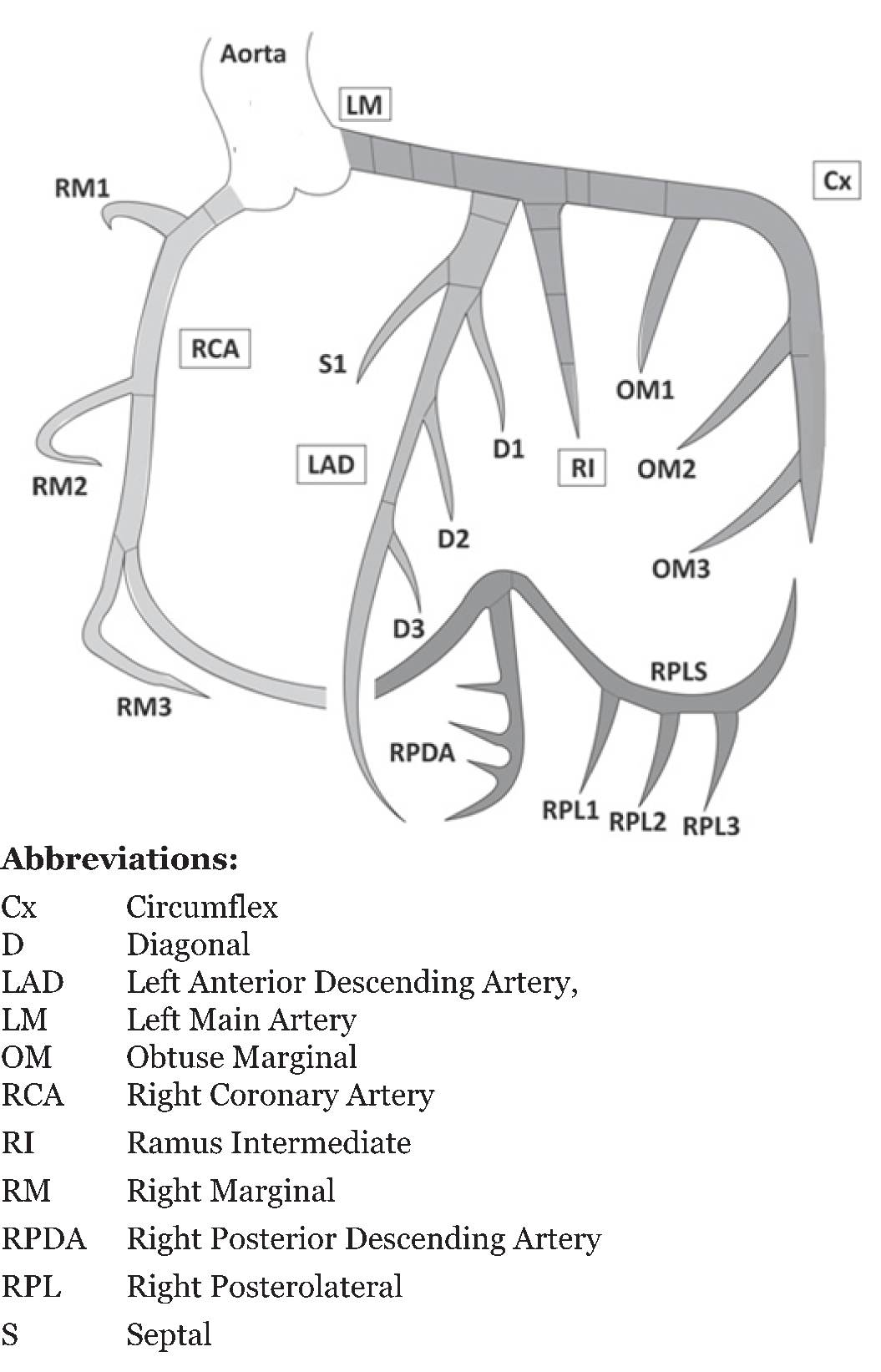
The study was conducted at the Department of Cardiology, Postgraduate Institute of Medical Education and Research (PGIMER), Chandigarh, India. A total of 321 patients who underwent coronary angiography for the evaluation of their symptoms and were found to have normal epicardial coronary arteries free of atheroma or stenosis were included in the study. Written Informed consent was obtained from each patient and the study protocol conforms to the ethical guidelines of the 1975 Declaration of Helsinki as reflected in a priori approval by the institution's human research committee. Procedure Selection Criteria for Target Vessels The digital angiograms were acquired on the GE digital cardio imaging (DCI) system and the images were transferred to a CD-ROM disk from the computer hard disk. The catheter used for angiography became the calibrator for the QCA system by employing an automated and operator independent edge detection technique in which the dimension of coronary artery was measured as a function of the catheter diameter. Computerized software analysis was used to calculate the vessel diameter in millimeter. Figure 1 represents various segments of coronary circulation that were analyzed. For the left coronary artery the segments were left main artery (LM), proximal left anterior descending artery (LAD) before origin of the first septal branch (S), mid left anterior descending artery between origin of first septal and first diagonal (D) branches, distal left anterior descending artery after the first diagonal artery, proximal circumflex (Cx) before origin of the first obtuse marginal (OM), distal circumflex after the origin of the obtuse marginal and the first obtuse marginal branch (OM). Similarly, for the right coronary artery the segments were proximal right coronary artery (RCA) before origin of first acute marginal, mid right coronary artery between first and second acute marginals, distal right coronary artery after the second acute marginal branch, posterior descending artery (RPPD) and the posterior left ventricular branch (PLV). The diameters of these coronary arteries were analyzed. Patients were also grouped as diabetics and non-diabetics and comparison of coronary artery diameters was done between these two groups. Statistical Analysis | ||||||
| ||||||
|
Results | ||||||
|
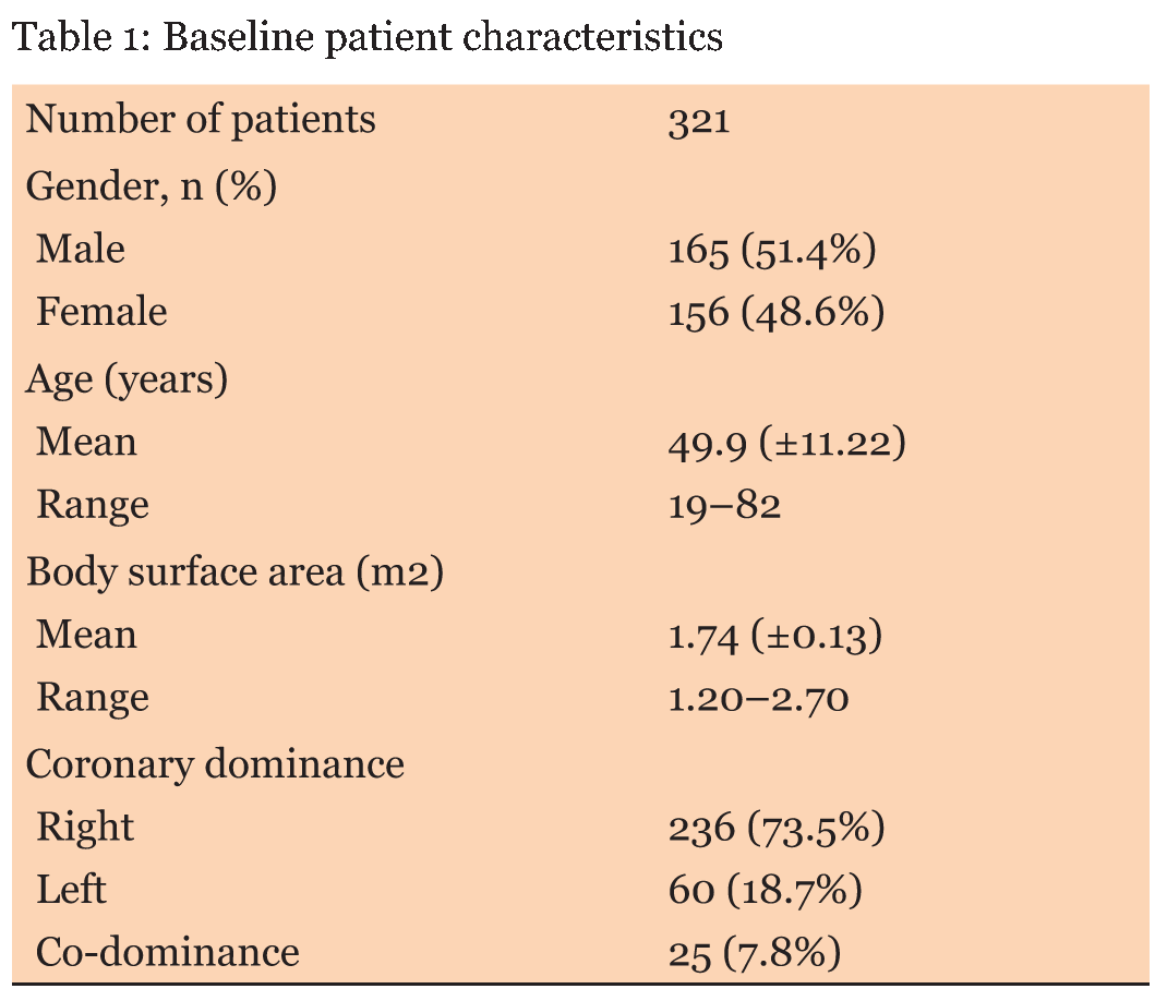
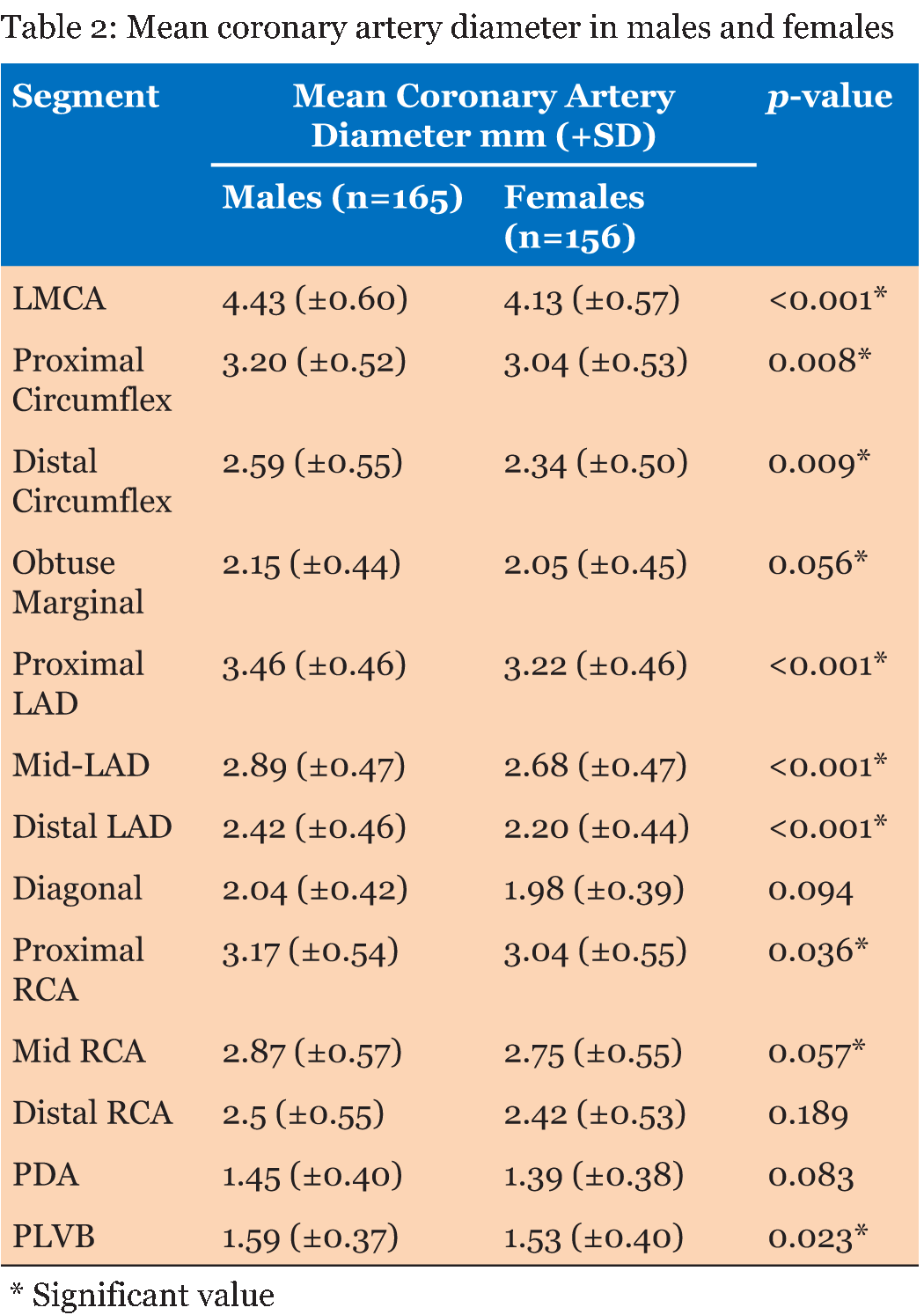
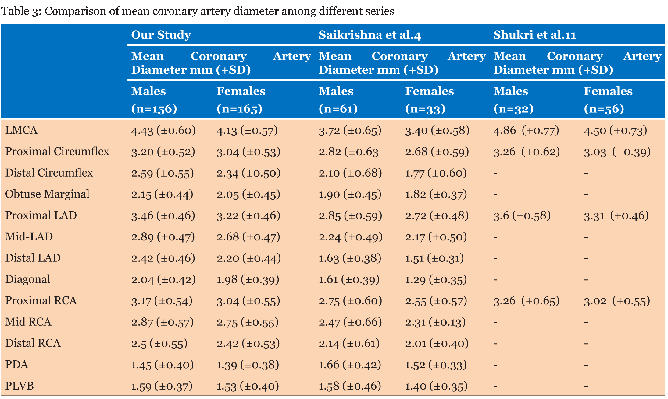
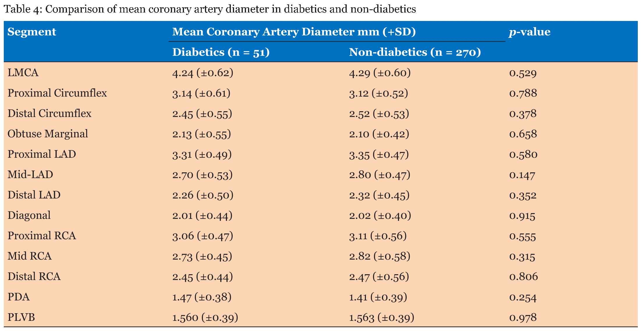
A total of 321 patients were included in this study between July 2013 and June 2014. There were 165 men and 156 women with a mean age of 49.9 years (±11.22) (range 30–67 years). Main baseline patient characteristics are enumerated in Table 1. Patients were matched for their age, clinical symptoms and risk factors like hypertension and diabetes. The mean age among males and females was 49.5 years (±12.5) and 50.4 years (±9.5) respectively. The mean body surface area among males was higher than females, which was statistically significant (p-value < 0.001). The mean coronary artery diameter was higher in males as compared to females Table 2 and the difference was statistically significant except for diagonal branch of left anterior descending artery, distal right coronary artery and posterior descending artery. The mean coronary diameter in our study is higher than the previously reported Indian population but lower than Iraqi Kurdish population Table 3. Our study also compared the mean coronary artery diameter in diabetics and non-diabetics sub-groups. 51 (15.9%) patients had diabetes whereas 270 (84.1%) patients were not diabetic. The mean age in diabetic and non-diabetic sub-group was 52.1 years (SD±8.8) and 49.5 years (SD±11.5), respectively. The diabetic patients had higher mean body surface area of 1.76 m2 (SD±0.09) as compared to 1.73 m2 (SD±0.14) in the non-diabetic patients. There was no statistically significant difference in either the age or BSA between the diabetics and non-diabetics (p value 0.13 and 0.15, respectively) patients. Although the mean coronary artery diameter in diabetic was lower than the non-diabetic patients, the difference was not statistically significant Table 4. | ||||||
| ||||||
| ||||||
| ||||||
|
| ||||||
| ||||||
|
Discussion
| ||||||
|
Various studies have reported that the dimensions of normal coronary arteries vary significantly among general population [3] [11] [12] [13]. These studies have correlated the genetic factors, age, sex, body surface area, heart weight and ethnic racial factors with the coronary artery anatomy [8] [12] [14] [15] [16][17][18]. Most of these studies have estimated the coronary artery size either in the patients undergoing coronary artery bypass grafting or in the post-mortem samples. However, visual interpretation of coronary dimensions without accurate quantification has been flawed with high inter-observer variability. Quantitative coronary angiography enables the operator to assess the size of the vessel before performing any intervention as well as obtaining the objective results of the intervention. This digital quantitative estimation of the coronary dimensions has been validated in various studies [18] [19] [20]. In the present study we measured the diameters of various segments of coronary artery and compared it among males versus females as well as diabetic versus non-diabetic patient population. The mean left main coronary artery diameter in our study was 4.28 mm (±0.6) and it was significantly higher in males as compared to female patients (p < 0.001). Similarly, the diameter of proximal circumflex, distal circumflex, obtuse marginal, proximal left anterior descending, mid- left anterior descending, distal left anterior descending, proximal right coronary artery, mid right coronary artery and posterior left ventricular branch of right coronary was also significantly higher in males as compared to female patients. These observations were in concordance with the results of previous studies by Saikrishna et al. [4], Dhawan and Bray [8], and Lip et al.[21]. The mean diameters of LMCA (4.28 mm), proximal circumflex (3.12 mm), proximal LAD (3.34 mm) and proximal RCA (3.11 mm) in our study was higher than one of the previous study from India by Saikrishna et al. [4]: LMCA (3.56 mm), proximal circumflex (2.75 mm), proximal LAD (2.79 mm) and proximal RCA (2.65 mm) but marginally lower than another study from India by Vikram et al. [22]: LMCA (4.3 mm), proximal circumflex (3.16 mm), proximal LAD (3.48 mm) and proximal RCA (3.15 mm) as well as a study in Iraqi Kurdish population by Shukri et al. [11]: LMCA (4.68 mm), proximal circumflex (3.15 mm), proximal LAD (3.46 mm) and proximal RCA (3.14 mm). Similarly, mean diameters in our study was higher to the Indo-Asians sub-group of the study conducted by Lip et al. [21]: LMCA (3.98 mm), proximal circumflex (3.01 mm), proximal LAD (3.22 mm) and proximal RCA (2.98 mm) but lower than the Caucasian sub-group: LMCA (4.44 mm), proximal circumflex (3.17 mm), proximal LAD (5.53 mm) and proximal RCA (3.35 mm). As coronary artery dimension correlates with the body weight, the marginally smaller mean coronary artery diameters in Indian population may be attributed to the lesser body weight of the Indian populations as compared to Caucasian and Iraqi Kurdish population. In the present study, we report a very high right sided dominancy of 73.5% as compared to 18.7% of left sided dominancy. These results are compatible with right sided dominancy ranging between 64.8% and 75% and left sided dominancy ranging from 6–8% reported for Indian, Caucasian and Iraqi Kurdish populations respectively. Our study also compared the mean coronary artery diameter in diabetic versus non-diabetic patients which to the best of our knowledge has not been compared till date. The result showed that mean diameter in diabetics was lower than non-diabetics. However, the difference was not statistically significant. Therefore, it can be interpreted that diabetics does not have narrower coronary arteries as shown in a previous study by Faridullah et al. [23]. | ||||||
|
Conclusion
| ||||||
|
Our study reports the largest single center data on coronary artery size in Indian population. The study included patients from 5–6 states of northern India, thereby having a diverse representation of the ethnic population as well as clinical presentation. Our study confirms the fact that coronary artery diameter is larger in males as compared to females. However, it contradicts the general belief that Indians have narrower coronary artery than their western counterpart. Also, there is no significant difference between the coronary artery diameter of diabetic patients as compared to non-diabetic patients. | ||||||
|
References
| ||||||
| ||||||

|
[HTML Abstract]
[PDF Full Text]
|
|
Author Contributions:
Saurabh Mehrotra – Substantial contributions to conception and design, Acquisition of data, Analysis and interpretation of data, Drafting the article, Revising it critically for important intellectual content, Final approval of the version to be published Shaadab Mohammed – Analysis and interpretation of data, Revising it critically for important intellectual content, Final approval of the version to be published Yashpaul Sharma – Analysis and interpretation of data, Revising it critically for important intellectual content, Final approval of the version to be published |
|
Guarantor of submission
The corresponding author is the guarantor of submission. |
|
Source of support
None |
|
Conflict of interest
Authors declare no conflict of interest. |
|
Copyright
© 2016 Saurabh Mehrotra et al. This article is distributed under the terms of Creative Commons Attribution License which permits unrestricted use, distribution and reproduction in any medium provided the original author(s) and original publisher are properly credited. Please see the copyright policy on the journal website for more information. |
|
|
|
About The Authors
| |||
| |||
| |||
| |||